Central Fringes of the Slit
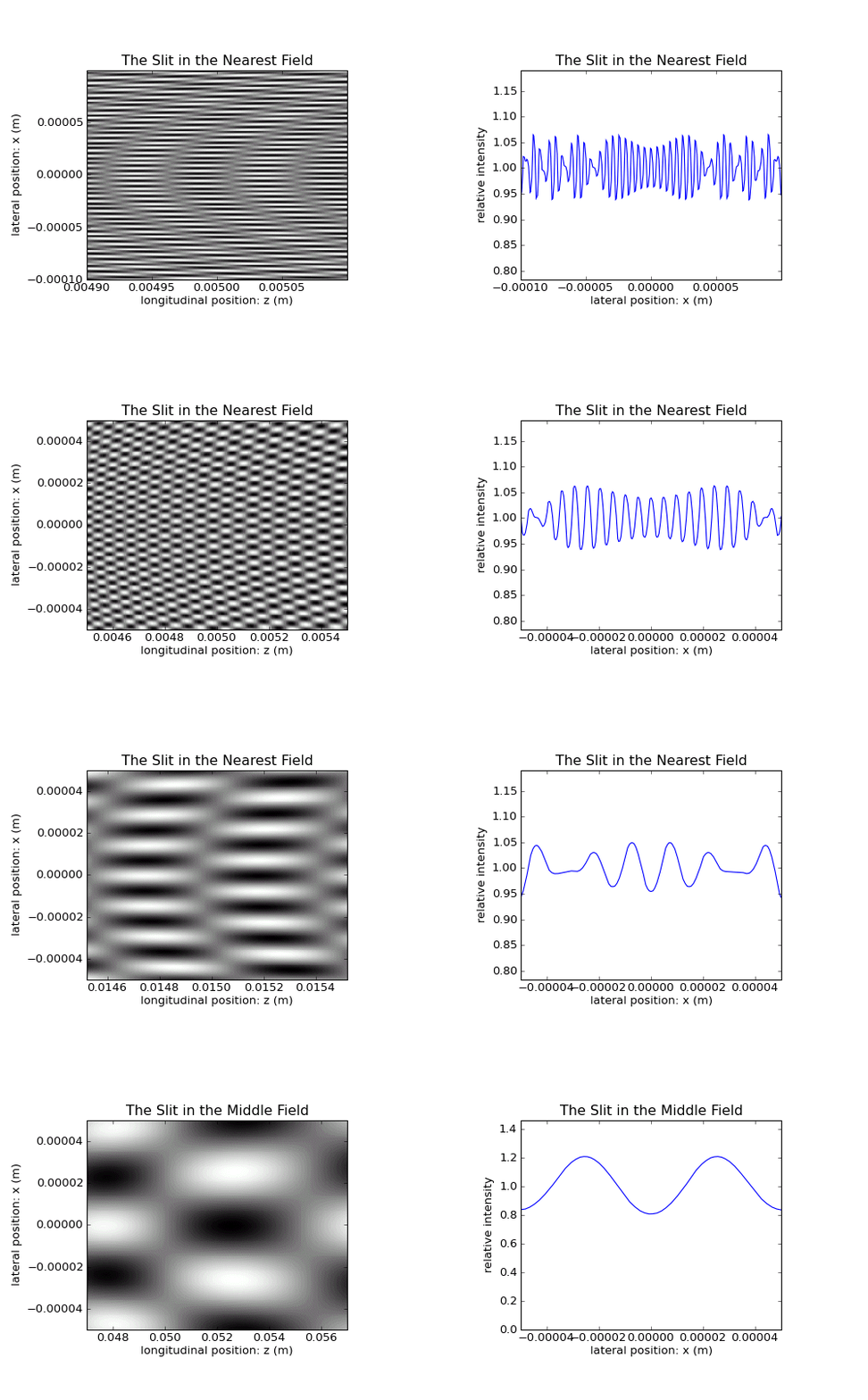
A closer look on the inner, early fringes of the slit.
The first picture is to scale, i.e., \(x\) and \(z\) have the same scaling factor. The picture is thus a magnification of the real observable pattern.
For the second and the following pictures, the scale is changed, such that the pattern becomes best visible. \(x\) and \(z\) have no longer have the same scaling factor. But for all pictures from the second on, this scale itself stays invariant, only the longitudinal \(z\) position is shifted to higher \(z\) on the optical axis.
One can discern, how the pattern stays in principle the same, and how it scales with growing \(z\).
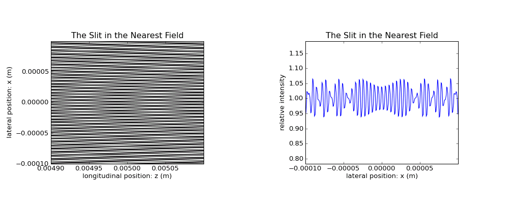
Tiny sublobes superimposed to the intensity near to 1.0 of the most central lobe can be easily discerned. The scale is proportional.
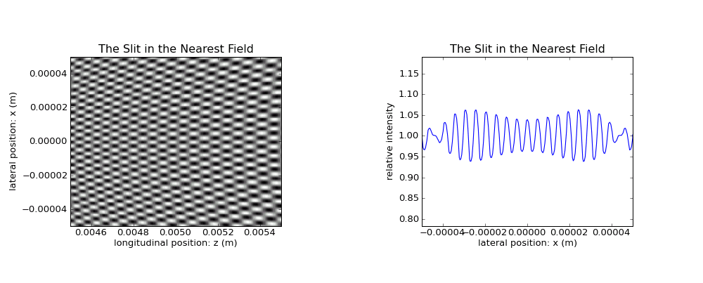
In a slightly different scale, their structure is better visible.
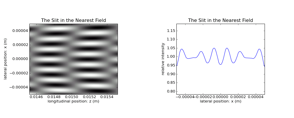
The size of the image is the same, but the \(z\) position is shifted.
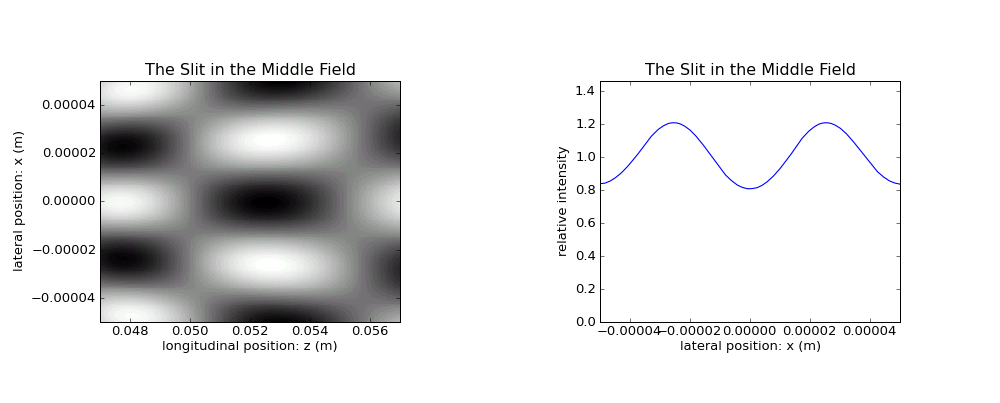
The size of the cells grows. Once they will will mature to the Fraunhofer diffraction pattern as described in the Overview Section